Politics, Research, Science, Uncategorized

Moderator Linsey Davis was wrong, and we’re missing the real question

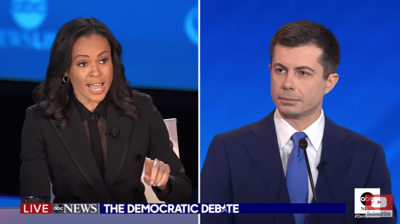
     Watch the first 2.5min of this exchange from last night’s Democratic Primary debate.
     I was unable to find a good fact check or even good coverage of this exchange – only hot takes and cheers from the far left that “Mayo Pete” finally got his comeuppance. However, the claims made by the moderator were pretty complex, so I dug into the data and did the analysis myself.
     First, Davis’s initial, very precisely worded claim that the ratio of black to white arrests for marijuana possession went up during Buttigieg’s mayorship was correct, but a bit misleading. While the ratio of black to white arrests did go up after Buttigieg took office, the number of possession arrests for both white and black people went down significantly during his tenure.
South Bend Analysis.jpg
     Would it have been better if black arrests had gone down even more than for whites so that the ratio was closer to one? Sure, but did he make things better? Of course he did.
     This data tells the story of the real left vs. far-left debate: Would black people in South Bend have been better off if Buttigieg had continued to arrest white people at the pre-2012 rate, making for a more equitable arrest ratio but arresting more people for a victimless crime? If you really understand these data and your answer is “yes”, I would suggest that you are are more interested in vengeance than justice or equality.
     That debate aside, I must turn my ire to moderator Lindsey Davis. Like many journalists, she started with an interesting issue but brought more heat than light.
     Davis pushed back against Buttigieg’s imprecise answer, and this is where she started making mistakes:
Davis: “How do you explain the increase in black arrests in South Bend under your leadership for marijuana possession?”
     This statement is flat out false. For the data we have, the rate of black arrests was significantly lower under Buttigieg and continued to fall for most of his administration. She probably meant the ratio of black to white arrests, but that is a completely different claim.
Buttigieg: “and again, the overall rate was lower than the national rate…”
Davis: “No, there was an increase. The year before you were in office, it was lower, once you became in office in 2012, that number went up. In 2018, the last number year that we have record for, that number was still up”
     Check out the data. Again, she’s probably meaning to say that the ratio of black to white arrests went up, which is true. However, what she is clearly saying, that the arrests of black people for possession went up, is wrong. Arrests of black people for possession went down while Buttigieg was mayor.
     There was a spike in 2018, but this is clearly an outlier and arrests that year went up for both white and black people and the ratio held steady.
     Buttigieg had an imprecise answer about his overall arrest rate, which isn’t quite the point I have been trying to make. Not unreasonably, he wasn’t completely up to speed on the data she was throwing at him. This is the actual “gotcha” journalism that I believe is unfair.
     As I watched this exchange live, what I mostly bumped on was their use of rates, ratios, and percentages to describe arrests in a city of only about 100,000 people. That’s too small of a city to describe in such terms as opposed to absolute numbers. You could bust one big college party and see the rate skyrocket. However, I’m sure that this is not an argument that Mayor Pete wants to make: highlighting that his city was so small that you probably can’t learn much about his governance from data like this.
     See what happens when journalists get sloppy… Warren jumped on Buttigieg not confronting the facts, the narrative about Pete’s performance gets distorted, we miss an important ideological debate, and already complex nomination process for the potential leader of the free world gets further confused.
     METHODS: The demographic data for annual arrests for possession of marijuana in South Bend, IN was downloaded from the FBI, the same data Davis was citing. I tried to get a baseline of how demographics in the city have changed, but couldn’t find a breakdown from year to year. Instead, I combined the data from the 2000 and 2010 census and another survey from 2017 to get a baseline of 60.25±3.60% white and 25.66±0.66% black residents of South Bend, IN. These standard errors were then propagated to give a general sense of the variation in baseline.


Standard
Science

Dinosaurs! Why did sauropods have long necks?

Sauropods are plant eating, long-necked dinosaurs, a group that includes the largest land animals ever to walk the earth. As much as sauropods permeate pop culture, their most striking feature, their long necks, remain mysterious. Why did nature select for such a long neck? Many assume that, like giraffes, the longer their neck, the higher food they could reach. However, this assumption is riddled with holes.

First, the blood pressure required to service a brain that high above the heart would push the limits of biology, perhaps requiring multiple hearts working in series or hardened veins to siphon blood up the neck. For comparison, healthy humans have a systolic blood pressure of about 120 torr. Giraffes: 180 torr, much higher to pump blood up a nearly two-meter neck. Sauropods, on the other hand, have to push blood through a vertical neck that is often over ten meters long, which would require blood pressure of approximately 600 torr! That presents a serious physics challenge that would require multiple hearts working in series or hardened veins, like plumbing pipes, to siphon blood up the neck. No evidence for such adaptations can be found in fossils or living animals.

Second, the torso of most sauropods is angled downward, with shorter front legs than back legs. Some scientists proposed that this simply shifts body weight toward the rear to facilitate rearing up on their hind limbs. However, we can rule out this unwieldy idea. The front limbs do not contain the micro-fractures that would be consistent with regularly returning to all fours.

Third, the sauropod neck does not appear to be very flexible. Computer modeling of its neck vertebra demonstrate that Diplodocus, if it strained to the limits of its skeleton, could not even raise its neck above parallel with the ground! These were clearly animals that browsed on low-lying plants. So, what were they reaching for?

Imagine what it’s like to be an animal that size. Weighing over a dozen tons, the stability of the ground they are standing on is a chief concern. Falling or getting stuck in the mud could be deadly. However, a long neck is an excellent way to get the head close to water or over treacherous ground while keeping the massive body on stable, dry land. Such a neck would have no need for flexibility, height, or ludicrous blood pressure. Perhaps long necks are simply an emergent property of being a land animal that large.

So that’s it, right? As compelling as this answer might seem, scientists don’t judge hypotheses by how satisfying they are. Ideas in science have power to the extent that they can be falsified and proven wrong. It is exceptionally difficult to falsify the idea that long necks evolved to span Paleozoic mud. There may be better solutions that no one has thought of yet. We must therefore tolerate ambiguity and accept only data-driven conclusions, because natural selection is far more creative than we.

Standard
Science

Tracking Bioengineered Bacteria Through The Body Using Ultrasound

Each of our hundreds of trillions of cells is as complex as a big city, and the mere shape of our internal organs reveals little about the true complexity of life. Unfortunately, you can’t “see” biochemistry, at least not with the naked eye. However, advances in molecular biology are literally illuminating the chemistry going on inside living tissues.

Scientists at the Shapiro Lab at Caltech have engineered bacterial cells that are exceptionally visible to ultrasound imaging. This technology could finally allow scientists to “watch” the biochemistry going on deep within living animals.

Since the 1990’s, there has been a fluorescent protein revolution in molecular biology. These fluorescent proteins are engineered to emit light from cells that express whatever gene scientists want to study. Unfortunately, unless your animal is translucent, they are not much use in illuminating the chemistry of internal organs. If you want to know what’s going on deep inside, you are limited to the static and specious snapshot of a dead and dissected animal.

GFP_hiir

Mouse engineered to express Green Fluorescent Protein (GFP)

Ultrasound imaging, on the other hand, is a great non-invasive technique to see inside a living body. This is the same technology that has us staring at amorphous black and white images trying to figure out how cute our future child will be. Ultrasound imaging uses echolocation, emitting sound waves (at a much higher pitch than we can hear) and recording their reflections from different internal tissues, turning them into a 3D image. Until now, ultrasound imaging only revealed internal structure, not the chemistry, gene expression, or types of cells in those deep tissues. However, the Shapiro Lab has engineered bacterial cells that specifically ‘light up’ under ultrasound.

Embryo_at_14_weeks_profile

Embryo at 14 weeks

How? Tiny balloons. Some bacteria in nature already make tiny gas-filled structures inside their cells to help regulate their buoyancy. Because of their size, these balloons happen to vibrate at the same frequency as the ultrasound wave, strongly reflecting the ultrasound signal. Obviously, these natural structures did not evolve with ultrasound technology in mind, so they had to be further engineered or tuned like tuning a guitar string or drum head. The Shapiro Lab did this by combining pieces of genes from different bacterial species to get the best ultrasound signal. Despite occupying roughly 10% of the cell’s volume and 1% of the cell’s mass, the balloons only marginally impaired the growth and movement of the bacteria.

An additional feature of these balloons is that they collapse when subjected to a suddenly strong pulse of ultrasound, like a glass shattered by an opera singer’s voice. The authors demonstrate that the balloons can be engineered to collapse at different pressures and frequencies, enabling different populations of cells to be distinguished within the same tissues, adding color to the gray ultrasound image.

As a proof of concept that these bacteria can be observed in live animals, the Shapiro Lab injected a chunk of gel that contained the engineered bacteria into the colon of a mouse. They used a species of bacteria that occurs naturally in the gut and is often used in humans to treat digestive disorders. Indeed, shape and location of the bacterial gel inside the mouse could be imaged with high resolution.

They were also able to image cancer tumors using this technology. Special strains of bacteria have already been engineered to bind to cancer tumors and even inhibit their growth. By giving these cancer-tracking bacteria the balloon-making genes, they successfully imaged a mouse tumor using ultrasound.

Bacteria can be easily engineered to stick to a variety of specific tissues, and have the potential to serve as ultrasound homing beacons for imaging various tissues in both research and medical therapy. Other applications include tracking the spread of newly introduced bacteria into our gut and the dynamics of our bacterial ecosystem. Many medical disorders are caused by imbalances in our gut bacteria and require the introduction of new bacteria into our body to correct the imbalance. Better understanding how and where these bacteria spread within us will better direct future treatments and innovations.

One of the Shapiro Lab’s most ambitious goals moving forward is engineering animal cells to manufacture these ultrasound balloons. However, can animal cells make this bacterial structure? Would the presence of these balloons affect the way animal cells behave? Would they stimulate an immune response? Can they be used in a therapeutic context? If these hurdles can be cleared, the impact of ultrasound markers like these could be an extraordinary new tool for research and medicine.

Standard